This technique can image over 10,000 genes at once
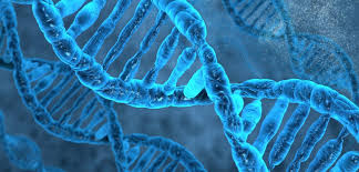 In a new breakthrough research, scientist have found a technique to image more than 10,000 genes at once within individual cells.
The new technique, dubbed intron seqFISH --sequential fluorescence in situ hybridisation--, is a major advance in being able to identify what goes on across the genome in hundreds of different cells at once where previously researchers could only image four to five genes at a time in cells with microscopy.
"Intron seqFISH can help identify cell types and also what the cells are going to do, in addition to giving us a look at the chromosome structure in the same cells," said Long Cai from Tianqiao and Chrissy Chen Institute for Neuroscience at California Institute of Technology.
The findings were detailed in the study published in the journal Cell.
In order for genetic instructions to be turned into an actual functioning protein, a process called transcription must first occur which often occurs in pulses, or "bursts".
First, a gene will be read and copied into a precursor messenger RNA, or pre-mRNA, like jotting a quick, rough draft. During the "editing" process, certain regions called introns are cut out of the pre-mRNA.
Using the newly developed intron seqFISH technique, each intron is labelled with a unique fluorescent barcode, enabling it to be seen with a microscope.
These introns then reveals which genes are currently turned on in individual cells, how strongly they are expressed, and where they are located. Because there are 10,421 introns, therefore 10,421 genes can be imaged at once.
Further, because introns stay where the gene is physically located, fluorescently imaging introns allows researchers to visualise where genes are located within the chromosome, the researchers explained.
Read more news: